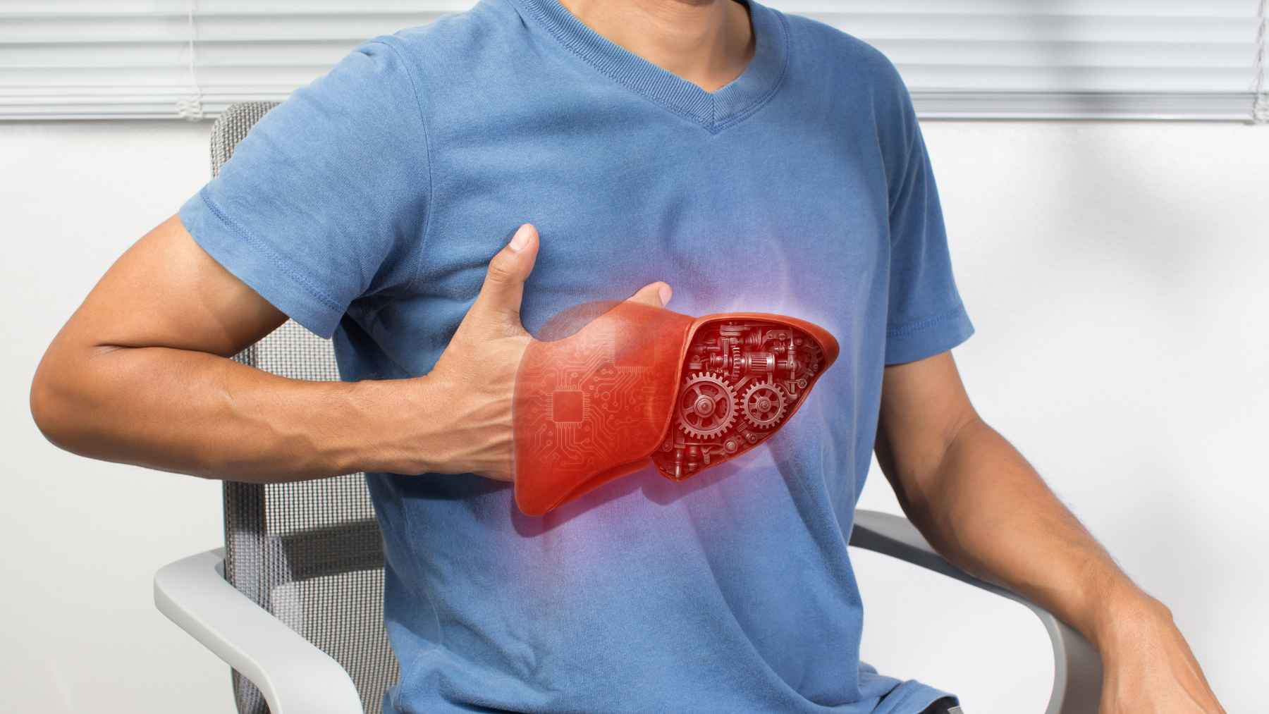
Millions of people live with fatty liver and never realize it. A new study finds that when colorectal cancer reaches the liver, this hidden condition may help tumors invade healthy tissue instead of remaining walled off.
Patients with the infiltrating growth pattern have five-year overall survival below 44%, compared with about 73% for the more contained pattern. The result does not show that fatty liver causes colorectal cancer, but it suggests the liver’s condition can influence how dangerous a metastasis becomes.
A condition many never notice
Fatty liver, also called liver steatosis, means excess fat has accumulated inside liver cells. It is often connected to obesity, type-2 diabetes, and other metabolic problems, yet many people feel completely well.
The National Institute of Diabetes and Digestive and Kidney Diseases estimates that about 24% of U.S. adults have a common form of fatty liver unrelated to heavy alcohol use–roughly one in four adults.
Colorectal cancer is also a major global threat. The World Health Organization recorded about 1.9 million new cases and more than 900,000 deaths in 2022, while several countries are reporting a growing burden among younger adults.
The growth pattern that matters
Metastasis happens when cancer cells leave the original tumor and establish new tumors elsewhere. The liver is the most common destination for colorectal cancer spread, and the way those new tumors grow can strongly affect the outlook.
Encapsulated metastases stay separated from nearby liver tissue by a border. “Replacement” metastases behave differently, weaving into healthy tissue and taking its place as they expand.
That second pattern is harder to treat, and no approved therapy specifically targets it. So what makes one patient develop it while another does not?
How liver fat changes cancer
The researchers found that extra fatty acids in a steatotic liver changed how colorectal cancer cells used fuel. The cells burned more fat, which helped stabilize MYC, a protein that acts like a growth switch in many cancers.
A more stable MYC protein pushed the cells to make more proline, an amino acid used to build proteins. Proline then supplied material for collagen, the tough substance that gives tissues structure.
Here, collagen worked like scaffolding. It helped cancer cells infiltrate the liver instead of remaining inside a clearly defined tumor border, linking a common metabolic condition to a specific high-risk form of cancer spread.
What the team tested
The team analyzed patient samples alongside mouse models, patient-derived tissue models, and organoids. Organoids are miniature tumors grown in a laboratory from patients’ cells, giving scientists a closer view of how real tumors may respond.
In the official release, senior author Sarah-Maria Fendt of the VIB-KU Leuven Center for Cancer Biology said a patient’s physiology is “not just a bystander, but an active determinant of disease progression.” First author Yiming Peng-Winkler said treatment must consider “not just the tumor, but also the environment it depends on.”
The researchers found that treatment-naive patients with fatty liver were more likely to have replacement metastases. “Treatment-naive” means they had not yet received cancer therapy that might change how the tumors looked or behaved.

Earlier clues were already there
A separate prospective study of 1,596 people with stage one through three colorectal cancer found that more liver fat was linked to higher recurrence and worse survival in colon cancer. The same clear association did not appear in rectal cancer, a reminder that colorectal cancer is not one uniform disease.
Earlier research also showed that proline metabolism can help cancer cells adapt during metastasis. The new work connects those clues in one chain involving liver fat, MYC, proline, collagen, and an aggressive growth pattern.
A possible treatment path
When the team blocked MYC, the enzyme needed to make proline, or collagen production, replacement metastases formed or grew less in preclinical models. That is encouraging, but these experiments were not a treatment trial in patients.
One MYC-targeting drug has already been tested in a small phase one trial involving 22 adults with advanced solid tumors. The trial mainly examined safety, so it cannot show that the drug will work for people with fatty liver and replacement metastases.
Still, liver fat could eventually serve as a biomarker, or measurable clue, to help doctors identify patients for future trials. Better selection could make those trials clearer and reduce exposure to treatments unlikely to help.
What the finding does not prove
The research does not show that fatty liver starts colorectal cancer. It also does not prove that reducing liver fat after diagnosis will stop metastases, reverse an existing growth pattern, or improve survival.
For now, the biggest shift is in how scientists view the disease. A tumor does not grow in isolation, and a common metabolic condition may change the ground beneath it in ways that matter for prognosis and treatment.
The full study was published in Nature.