A new Alzheimer’s study suggests that the brain’s own immune cells may play a major role in the loss of deep, restorative sleep. In mice with amyloid plaques, temporarily removing most of those cells restored more than two hours of sleep a day even though the plaques remained.
The finding shifts attention away from plaques alone and toward the inflammation surrounding them. What if the brain is being kept awake not only by damage, but also by an immune response that never seems to switch off?
Brain immune cells may be keeping the brain awake
Microglia are immune cells that live inside the brain and respond to injury, infection, and abnormal protein buildup. They normally protect brain tissue, but the new work suggests they can become overactive when sticky amyloid plaques begin to accumulate.
Shannon L. Macauley, an associate professor of physiology at the University of Kentucky College of Medicine, led the research with Nicholas J. Constantino as first author. “Basically, we showed that it is not the plaques themselves, nor solely dysfunctional neurons, causing the lack of sleep, but rather the microglia,” she said.
The team compared the process to a sprinkler system flooding an entire house while trying to put out a small kitchen fire. In practical terms, a protective response may become so widespread that it keeps the brain in a heightened state when it should be resting.
Researchers tracked sleep as plaques developed
The scientists compared mice genetically prone to developing amyloid plaques with “wild-type” mice that aged normally. They examined the animals at six months, when plaques first appeared, and at 18 months, when the disease model was more advanced.
Small head-mounted devices recorded brain waves and muscle activity, allowing the researchers to identify waking, deep nonrapid eye movement sleep, and rapid eye movement sleep associated with dreaming.
Light-sheet microscopy also produced three-dimensional maps showing plaques and immune cells throughout the brain.
Early plaques caused a lasting sleep deficit
One of the most unexpected findings was that worsening plaque buildup did not produce a matching decline in sleep. By 18 months, the mice had more than twice the plaque burden seen at six months, yet their sleep disruption and abnormal brain activity had not become substantially worse.
“I expected that as plaque burden became more severe, sleep disruption would also worsen,” the first author said. Instead, the results pointed to a “ceiling effect,” meaning the first wave of immune activation may have been enough to establish the problem.
That changes the timeline researchers may need to watch. The trouble may begin early, when plaques first appear and microglia launch an intense response, rather than only after large amounts of amyloid have accumulated.
Deep restorative sleep was hit hardest
The study separated changes linked to normal aging from those associated with amyloid disease. Aging mainly reduced rapid eye movement sleep, while amyloid pathology selectively reduced nonrapid eye movement sleep, the deeper stage that supports physical recovery, learning, memory, and the brain’s nightly waste-clearing activity.
Anyone who has stumbled through a morning after poor sleep knows how quickly attention and thinking can suffer. With Alzheimer’s-related disease, repeated loss of restorative sleep may be more serious because it could weaken waste removal and create a cycle of further damage and more sleep disruption.
“That restorative sleep is super important for physical repair, learning and memory and washing out the toxins of the day,” the lead researcher explained. The mouse findings do not prove that the same mechanism works identically in people, but they give scientists a focused question to test.
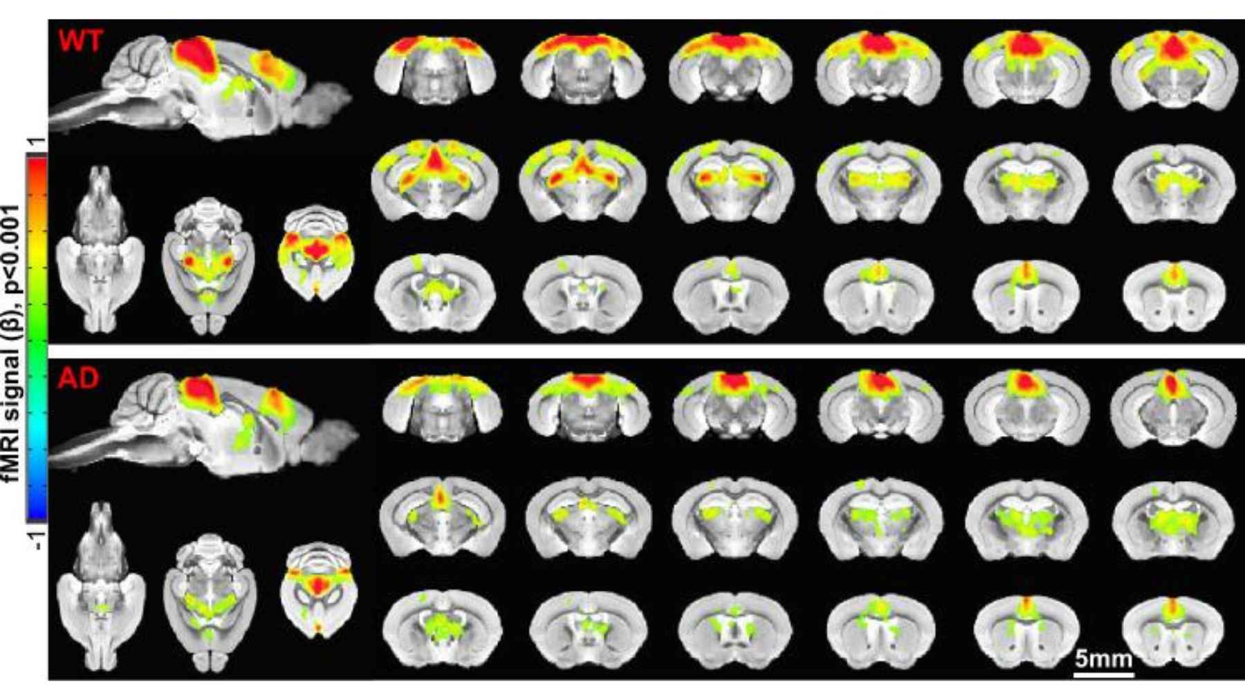
Brain imaging compares healthy mice with Alzheimer’s disease mice, highlighting differences in activity across several brain regions.
More than two hours of sleep returned
To find out whether microglia were causing the disruption, the team gave the mice pexidartinib, also known as PLX3397, for 14 days. The drug blocks a survival pathway used by microglia and temporarily removed about 87% of the brain’s immune cells.
After that depletion, mice with Alzheimer’s-related pathology gained more than two hours of sleep a day. Their restorative nonrapid eye movement periods also became longer, giving them more opportunities to enter dreaming sleep linked to memory formation.
The amyloid plaques did not decline. That is the key point. The result suggests that inflammation around plaques may be a reversible contributor to sleep loss, separate from the plaques themselves.
The next goal is calming microglia safely
The experiment does not mean that removing microglia is a ready treatment for people. These cells perform essential protective jobs, and the drug was used as a research tool to test cause and effect in mice.
The laboratory at the Sanders-Brown Center on Aging is now studying whether microglia can be calmed instead of eliminated.
Researchers are examining existing medicines, including the diabetes drug metformin and stiripentol, to see whether changing how the cells use energy can reduce overactivity, but no benefit for Alzheimer’s patients has been established.
The team also identified electrical brain patterns that may help distinguish Alzheimer’s-related changes from normal aging. Portable EEG systems could eventually offer a lower-cost way to monitor people at home or in local clinics, although that possibility still needs human testing.
The main study has been published in Alzheimer’s & Dementia.