
Could a worn-out knee repair its own smooth lining instead of being replaced with metal and plastic? A new preclinical study found that blocking one aging-linked enzyme caused old mice to rebuild articular cartilage, the slick layer that helps joints glide with little friction.
Cartilage taken from people undergoing knee replacement also began showing repair signals after treatment in the lab.
The finding offers a possible new route for osteoarthritis, but it does not prove that a pill or injection can regrow cartilage inside a living human joint, and it does not mean orthopedic surgeons are about to lose their jobs.
Why osteoarthritis is hard to reverse
Osteoarthritis is the most common form of arthritis and develops as cartilage and other joint tissues break down over time. The World Health Organization estimated that 528 million people were living with it in 2019, with knees affected most often.
That damage can turn ordinary things, such as climbing stairs or getting out of a car, into a painful calculation. Current care includes exercise, weight management, pain relievers, anti-inflammatory drugs, and injections, while advanced disease may require joint replacement.
Those options can reduce symptoms and restore mobility, but they do not reliably rebuild the original smooth cartilage. Researchers are still looking for an approved medicine that can slow, stop, or reverse the structural damage, which is why this result has drawn attention.
The aging enzyme behind the result
The target is 15-PGDH, an enzyme that becomes more abundant in several tissues with age. The researchers call it a “gerozyme,” meaning an aging-linked enzyme that helps drive the gradual loss of tissue function.
In old mouse knee cartilage, 15-PGDH levels were about twice those found in young animals. The enzyme breaks down prostaglandin E2, a natural chemical signal involved in repair, so blocking it leaves more of that signal available.
The idea has a longer history. Research published in 2015 showed that inhibiting 15-PGDH boosted regeneration in several mouse tissues, giving scientists a reason to test whether the same pathway could help stubborn joint cartilage.
Cartilage cells reset without stem cells
The work came from Stanford Medicine, with contributions from Sanford Burnham Prebys Medical Discovery Institute, and was led by Mamta Singla and Yu Xin Wang. Senior author Nidhi Bhutani said, “Cartilage regeneration to such an extent in aged mice took us by surprise.”
Researchers gave old mice a small-molecule inhibitor either throughout the body or directly into the knee. In both cases, thinned cartilage became thicker across the joint surface, and tests identified it as functional hyaline cartilage, the smooth type joints need, rather than the tougher, less flexible repair tissue known as fibrocartilage.
The repair did not depend on stem cells. Existing cartilage cells changed which genes they used and shifted toward a younger pattern, while the share linked with healthy articular cartilage rose from about 22% to 42%.
Helen Blau, the other senior author and director of the Baxter Laboratory for Stem Cell Biology, said, “We were looking for stem cells, but they are clearly not involved.”
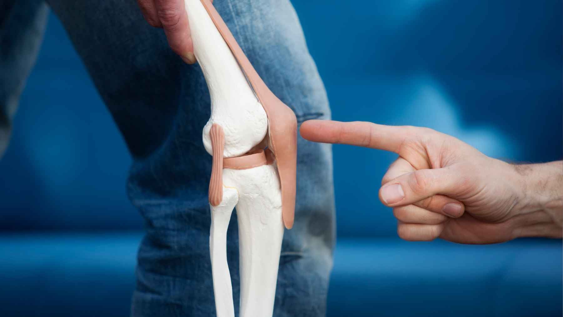
A knee joint model illustrates the cartilage affected by osteoarthritis, the focus of new research exploring regeneration by blocking an aging-linked protein.
Injury and human tissue tests
The team also created a knee injury in mice similar to an anterior cruciate ligament tear. Inhibitor injections given twice a week for four weeks sharply reduced later joint damage, and treated animals moved more normally and placed more weight on the injured leg.
About half of people with this common sports injury develop osteoarthritis in the affected joint within roughly 15 years.
The human test used cartilage from 11 patients ages 55 to 75 who were undergoing total knee replacement. After one week outside the body, treated samples showed fewer destructive cartilage cells, less activity from inflammation and breakdown genes, and early production of new articular cartilage.
Still, tissue in a laboratory dish cannot reveal whether a person would feel less pain, walk farther, avoid side effects, or keep the new cartilage for years. That is the line future clinical trials must cross.
Why joint replacement is not over
No human cartilage trial has yet reported that this approach repairs an arthritic knee or hip. Mouse joints carry weight differently, and researchers must determine the right dose, treatment length, delivery method, and long-term effects of changing prostaglandin signaling.
An oral inhibitor of the same target called MF-300, developed by Epirium Bio, completed Phase 1 testing in 100 healthy adults.
The company’s Phase 1 results reported no serious adverse events after single doses or five days of daily dosing, and the developer now lists the drug in a Phase 2b study for age-related muscle weakness. That is useful safety information, but it is not proof of cartilage repair.
Human osteoarthritis studies will need to measure pain, movement, imaging changes, durability, and whether treatment can truly delay or prevent surgery. The official release also disclosed related patent applications, a license to the drug developer, and a financial interest held by one senior author.
For now, joint replacement remains a well-established option for end-stage disease, while this enzyme blocker remains a promising but unproven regenerative strategy.
The official study has been published in Science.